Obfuscated Volume Rendering
Jia-kai Chou and Chuan-Kai Yang
Computer Graphics & Multimedia Lab., NTUST
| Abstract |
Analyzing and processing various data types
in a privacy-preserving perspective has been researched in
many disciplines; however, such an issue draws very limited
attention in the research field of scientific visualization. We
wondered if it is possible to delegate the rendering of a volume
data set to a remote server(s) while still being able to
preserve its privacy to certain extent. This paper presents
a block-based volume data transformation algorithm that
obfuscates a volume data set so as to reduce the user?�s privacy
concern when the volume data set is to be uploaded to
a remote server. In addition, a privacy-aware transfer function
adjustment is proposed so that not only the privacy is
protected during the rendering process, but also the computational
loading could be leveraged to the server side as
much as possible. Experimental results show that the proposed
method yields visually satisfactory results compared
with a normal direct volume rendering approach. Moreover,
the decrease of the rendering efficiency caused by the proposed
method is still controlledwithin an acceptable range.A
case study proves that the proposed approach can be adopted
in practice. This work explores the possibility of rendering a
volume data set through remote server(s) while the privacy
of data is still maintained.
|
[Paper]
[Supplementary Results] |
|
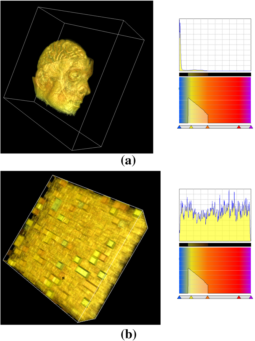
Figure 1: (a) A rendering result of the ?�MRBrain??data set (256 ? 256 ?
109 voxels) and its corresponding voxel value histogram and a user-assigned
transfer function. (b) The same transfer function applied to the
transformed ?�MRBrain??data set. |
|
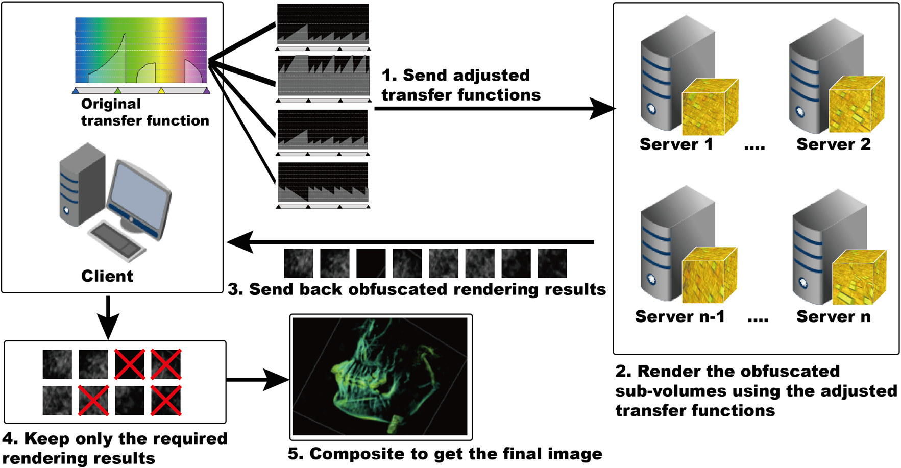
Figure 2: The overview of the flow of the remote rendering process. 1) The user-assigned transfer function is used to generate several adjusted transfer functions, which are then sent to the server side. 2) The server
side renders the corresponding sub-volumes according to the given
adjusted transfer functions. 3) The server side sends back the obfuscated
rendering results to the client side. 4) The client side keeps only the
required rendering results while discarding the rest. 5 )The client side
performs the inverse permutation to set the rendering results of each
sub-volume in the desired order, and then composites them altogether
to get the final image. |
|
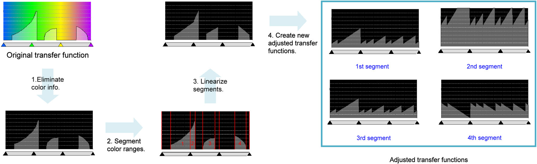
Figure 3: A step-by-step illustration of the proposed transfer function
adjustment. For a transfer function, we first remove its color information.
Then, the color ranges are divided into segments according to the
assigned alpha values. The segmentation boundaries and the segments
with non-zero alpha values are highlighted in red. After that, the segments
are linearized into pieces of straight lines. Finally, each segment
with non-zero alpha values is used to generate a new adjusted transfer
function with equalized slope and intercept in all segments. The corresponding
segments in the original transfer function that are used for
creating the adjusted transfer functions are marked in blue. |
|
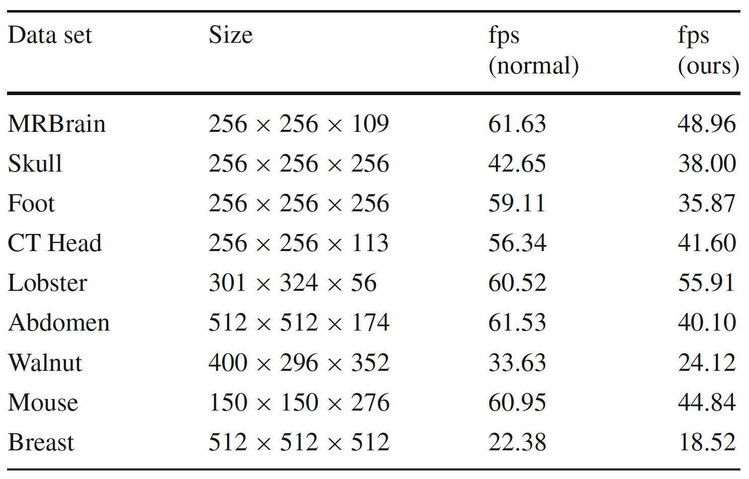
The rates of frame per second (fps) required while exploring
the volume data sets are measured to compare the rendering efficiency
between the proposed method and the normal volume rendering technique. |
Case Study
|
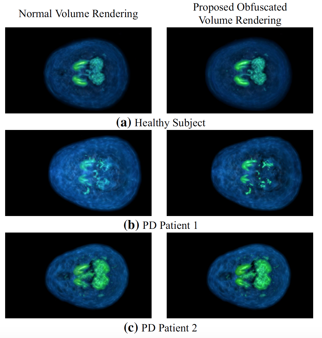
Comparing the volume rendering results of human brains
between a healthy subject (a) and two PD patients (b) and (c). In
addition, each human brain is rendered by both normal direct volume
rendering technique (shown on the left column) and the proposed obfuscated
volume rendering approach (shown on the right column). It shows
that the proposed method yields comparable rendering images with the
normal rendering technique. As can be seen in a and b, it is obvious
that the shape and silhouette of bilateral caudate, NAc, APu and PPu are
more symmetric and visible in the health subject (a) than in PD Patient
1 (b). However, when a PD patient is with mild symptoms (PD Patient
2), the reduction of VMAT2 may not be obvious enough for one to tell
whether the subject suffers from PDor not. In such a case, it may require
us to extract a specific slice(s) of the data for further investigation. |
|
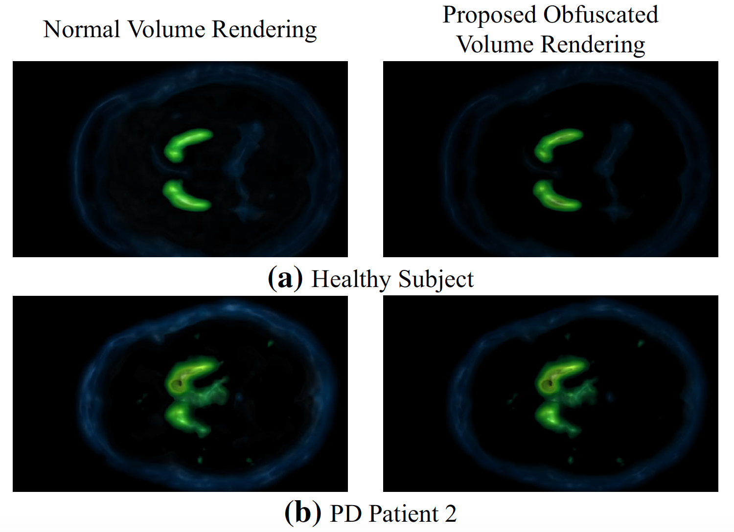
Approximating the rendering of one single slice or any specifically
chosen slices by rendering and compositing nearby sub-volumes. In this example, only the sub-volumes such as bilateral caudate, APu
and PPu reside in are involved. It can be seen that the contralateral PPu
gradually reduces in PD patient 2, while the features in the healthy subject
are more symmetric and clearly visible. Moreover, we also show
that the images rendered by the proposed approach (shown on the right
column) are close enough to the results generated by a normal rendering
method without affecting the visual assessment. |